New Digital Palm-Size Black / White Ultrasound Scanner For Human Use
Product Information
1.1 Characteristic
The high resolution Ultrasonic Diagnosis Equipment, controlled by computer and equipped with digital screening converter (DSC), is characterized by its clear image, stable performance and high resolution with adopting variable aperture, multi-segment dynamic focus, large dynamic band low-noise pre-amplifier, logarithmic compression, TGC control, dynamic filter, edge enhancement and frame-related technologies etc.
A. Use advanced production technology, multi-acoustic matching, frequency bandwidth, high sensitivity.
B. Full-digital transmission techniques, the received signal focus, good resolution
C. Rich software package. It is more suitable to users’ requirements.
D. Use switching power supply, power adaptability
E. Use high-speed CMOS integrated circuits and 1.5V low voltage deviceds, low energy consumption, no hear after continuous work and high reliability.
F. The circuit boards from the machine use surface mount technology, small size, light weight, beautiful appearance
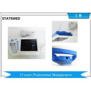
1. 2 Composition
The mode is the palm ultrasound scanner including mainframe, probe, screen and power adapter. Scanning mode is electron convex array and electron liner array.
Standard configuration: 3.5MHz R60 convex probe (80 elements)
Optional accessories: 6.5 MHz R10 tans vaginal probe (80elements),
6.5MHz linear array probe(80 elements)
Medical Image workstation software, printer, etc.
1. 3 Application
The equipment is designed as a multiple function ultrasonic product for your general and special clinical application of the check of pregnant and intrauterine contraceptive ring in family planning, gynecology and obstetrics, etc, to diagnose human liver, gallbladder, spleen, kidney, pancreas, bladder, womb and fetus.
1. 4 Contraindication
The product cannot apply to the burned, scald or injured surface texture of human body.
When choose trans-vaginal probe, it can not be used for the following patients.
Vaginal inflammation, such as trichnomonas vaginitis, fungal vaginitis, venereal disease, unmarried, viginal malformations, menstrual period, postmenopausal vaginal atrophy, transvaginal ultrasound difficulties, vaginal bleeding, patients with placenta previa.
Technique specifications
2. 1 Technical Parameters
Scanning mode: Electron convex array scanning,
Electron linear array scanning
Electron convex array
Five display format: B, B+B, 4B, B+M, M
Image magnification: ×1.0, ×1.2, ×1.3, ×1.5, ×1.6, ×1.8, ×2.0
L-zoom: real-time partial amplification
Depth of detection: ≥180mm
Image grey scale: 256 levels; 4-stageγcorrection
Frame: 3-stage frame function
Focus: Dynamic digital electronic focus point by point
Gain control: Full gain and 8-segment TGC
Probe Conversion: probe automatic identification
Frequency for Convex : 2.0MHz, 2.5 MHz, 3.0 MHz, 3.5 MHz, 4.0 MHz, 4.5 MHz, 5.0 MHz
Note: Hospital name, age, sex; organ automatic annotation function; Partial and full-screen character annotation function.
Display: English/Chinese conversion function; Fixed characters, real-time colock, calendar display.
Human Body Mark : 15 kinds
Puncture function: puncture guide line displayed in B mode.
Measurement function: distance, CRL, GS, BPD, FL, AC, HC, APTD, APD. Image display: image freeze, real-time, up/down, left/right, black/white reverse.
Cine loop: 512-frame cine loop and storage.
Image storage: store 16 thousand image by USB flash.
Printing function: connect with laser printer or video printer
Power saving function
Output interface: USB interface, standard mini port (and trapezoidal interface), video printer interface
Print: machine can connect any video printers directly.
Equipment for the injection shell structure. The host uses non-frequency transformer switching power supply, and adopt to the programmable devices and surface mount technology (SMT). The whole machine with high degree of integration, excellent performance.
2. 2 Main Technical Features
2. 2. 1 performance of standard probe
2. 2.1.1 R=60 the center frequency of convex array probe: 3.5Mhz
2. 2. 1. 2 investigation depth: >=180mm
2. 2. 1. 3 Vertical resolution:<=1mm(depth<=130mm)
<=2mm(130mm<depth<=170mm)
2. 2. 1.4 Lateral resolution:<=2mm(depth<=130mm)
<=3mm(130mm<depth<=160mm)
2.2.1.5 Geometric position accuracy: Portrait<=5%; cross wise<=10%;
Dead zone: <=4mm
Slice thickness : <=5mm
Pupil deviation:<=+-20%
M mode display error: <=5%
Main configure probe 3.5Mhz(R=60) the frequency of convex array probe: 2.0Mhz, 2.5Mhz, 3.5Mhz, 3.5Mhz, 4.0Mhz, 4.5Mhz, 5.0Mhz. The working frequency performance requirements listed in Table 1;
Table 1: B ultrasonic diagnostic performance requirements file
| performance | Probe style and standard frequency |
| Standard frequency of probe | 3.5 MHz | 6.5MHz |
| Random error of frequency | Deviation of Sound frequency and standard frequency should ≤±15% |
| Probe | linear, R≥60mmconvex | linear(linear), R≥60mm convex | R<60mm convex (transvaginal) |
| Investigation depth,mm | ≥180 | ≥70 | ≥60 |
| Axial resolution,mm | ≤2 (depth≤80) ≤3 (80<depth≤130) | ≤1 (depth≤50) | ≤1 (depth≤40) |
| Lateral resolution,mm | ≤3 (depth≤80) ≤4 (80<depth≤130) | ≤2 (depth≤40) | ≤2 (depth≤30) |
| Geometric position accuracy,% | crosswise≤10 Length wises≤5 | crosswise≤5 Length wises≤5 | crosswise≤10 Length wises≤5 |
| Dead zone,mm | ≤4 | ≤3 | ≤4 |
| Slice thickness,mm | ≤9 | ≤5 | ≤5 |
| Pupil error,% | ≤±20 | ≤±20 | ≤±20 |
| M mode time error,% | ≤5 | -- | -- |
| M mode distance error,% | ≤5 | -- | -- |
2. 2. 2 frequency of optional probes and performance requirements
2. 2. 2. 1 6.5Mhz (r=10mm) convex transvaginal: 4.0Mhz/6.0Mhz/6.5Mhz/7.5Mhz/8.0Mhz. Pls see the table 1 about the different frequency performance requirements
2. 2. 3 power supply voltage
Power supply AC220V+-22V, machine can work well
2. 2. 4 work time should >=8H
2. 2. 5 acoustic output pere maters
Response to acoustic output parameters of the device according to GB / T 16846-2008 requirements for testing, does not meet the conditions from published should be background information technical manuals, operating instructions, etc. to be published (see Appendix I)
2. 2. 6 diagnostic device according the function configure as
Medical imaging work station, which should be satisfy with requirements.
2. 3 Configuration
2. 3. 1 monitor: 7inches high resolution LED monitor;
2. 3. 0 dimension: 140mm*70mm*10mm
2. 3. 3 weight: 1Kg;
2. 3. 4 probe socket: 1 pc;
2. 3. 5 standard configuration: see packing list